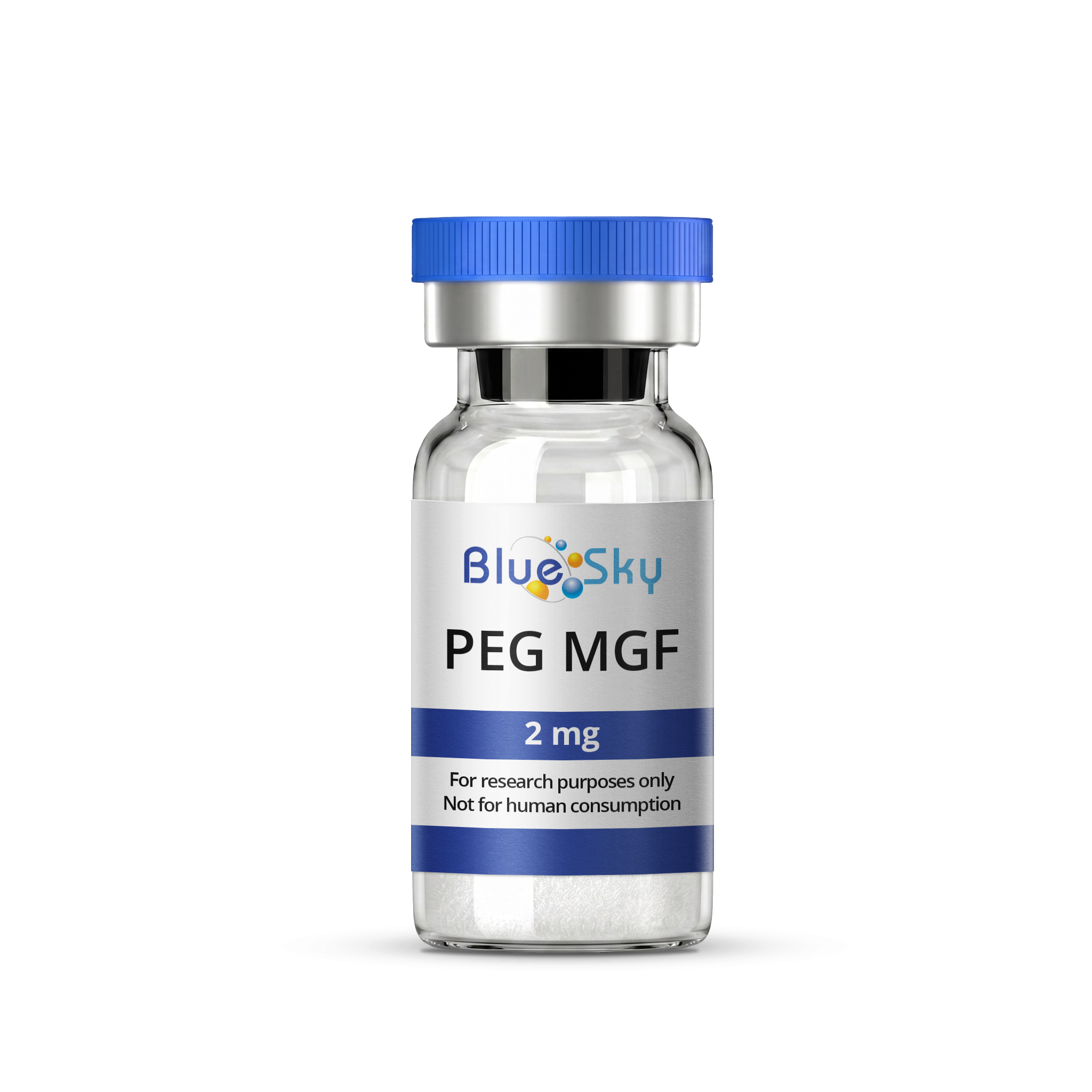
PEG MGF 2mg

- Free Priority Shipping
- Orders over $75



Buy PEG MGF 2 mg :
| Unit Size | 2 mg/vial |
| Unit Quantity | 1 vial |
| Purity (Mass Spectrometry and UV) | 99.64% |
| Sequence | PEG-Suc-Tyr-Gln-Pro-Pro-Ser-Thr-Asn-Lys-Asn-Thr-Lys-Ser-Gln-Arg-Arg-Lys-Gly-Ser-Thr-Phe-Glu-Glu-His-Lys-NH2 |
| Molecular Formula (MGF core peptide, pre-PEGylation) | C121H200N42O39 |
| Appearance | Lyophilized White Powder |
| Source | Chemical Synthesis |
| Storage |
Lyophilized PEG MGF is stable at room Temperature for 90 days, however it is best to store in a freeze below - 8c for any extended period of time. |
| Terms | The products we offer are intended for laboratory research use only. Please familiarize yourself with our terms of service prior to ordering. |
PEG MGF 2mg
View Research Overview & References
PEG MGF is a synthetic analog of mechano-growth factor (MGF), a splice variant of insulin-like growth factor-1 (IGF-1).
PEG MGF is created through a process called PEGylation, where polyethylene glycol (PEG) is attached to the molecule.1 This modification is studied for its relevance to the peptide's stability in laboratory assay conditions, since unmodified MGF is understood to degrade relatively quickly once introduced into cell culture media.
Human Mesenchymal Stem Cell Migration Research
In vitro studies using biomaterial microrods coated with MGF and PEG, designed to mimic cardiac tissue stiffness, found increased migration of human mesenchymal stem cells (hMSCs) toward the MGF-loaded microrods in this cell-based system.2
Cartilage Cell Migration Considerations
MGF is also studied in laboratory research for its association with chondrocyte migration behavior, a topic of interest given the growth plate's role in cartilage tissue development.
PEGylation Chemistry and Molecular Design
PEG MGF's structure combines the 24-residue MGF-E domain peptide sequence with a polyethylene glycol chain attached via a succinyl linker at the N-terminus.
Because polyethylene glycol is a polydisperse polymer rather than a single discrete molecule, the complete PEGylated conjugate does not have one fixed molecular formula the way the unmodified peptide does; its total mass instead depends on the specific PEG chain length used during synthesis.
Researchers comparing analytical data across suppliers are encouraged to confirm which PEG chain length was used, since this determines the conjugate's expected mass.
Researchers studying PEGylated peptides note that this modification increases the molecule's hydrodynamic radius substantially beyond what its peptide backbone alone would suggest, a size increase studied for its effect on the molecule's diffusion behavior and its retention characteristics in size-exclusion chromatography relative to the unmodified peptide.
Analytical Purity Verification
Purity verification for PEGylated research peptides typically requires confirming both the peptide sequence and the successful attachment of the PEG group, since incomplete PEGylation can leave a mixture of modified and unmodified peptide with different mass and chromatographic behavior.
Researchers reviewing a certificate of analysis for PEG MGF are encouraged to confirm that analytical data addresses PEGylation efficiency specifically, not just overall peptide purity.
Storage and Handling in the Laboratory Setting
Lyophilized PEG MGF is stored under sub-zero freezer conditions to preserve integrity prior to use, consistent with standard handling practices for PEGylated research peptides.
Important Notice
PEG MGF is intended exclusively for in vitro laboratory research and is not for human consumption, diagnostic use, or therapeutic use. These findings do not establish safety, efficacy, or suitability for any human application, and have not been evaluated by the FDA or any regulatory agency.
References
1. Veronese FM, Mero A. The impact of PEGylation on biological therapies. BioDrugs. 2008;22(5):315-29.
2. Doroudian G, Pinney J, Ayala P, Los T, Desai TA, Russell B. Sustained delivery of MGF peptide from microrods attracts stem cells and reduces apoptosis of myocytes. Biomed Microdevices. 2014 Oct;16(5):705-15.



 Login
Login